Plants have always been silent storytellers. They don’t speak, yet every wilted leaf, every subtle change in color, every shimmer under sunlight carries a message about what’s happening inside them. For centuries, plant physiologists have relied on their eyes and instruments to decode these messages counting stomata, measuring chlorophyll, or tracking gas exchange. But today, a quiet revolution is changing the way we “see” plants. Thanks to hyperspectral imaging, scientists can now detect stress in plants long before the naked eye notices a single yellow patch.
This technology is fast becoming the new eye of plant physiology an eye that doesn’t just see green and yellow, but hundreds of invisible shades that tell us how a plant truly feels.
What Exactly Is Hyperspectral Imaging?
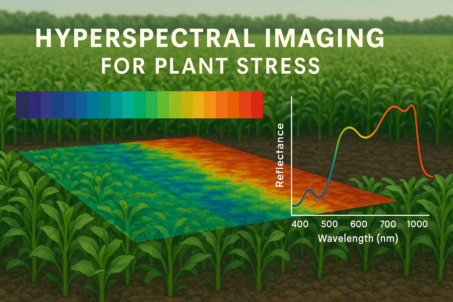
To understand hyperspectral imaging, imagine how we see color. The human eye detects just three broad bands of light red, green, and blue. Every color we perceive is a mixture of these three. But plants reflect and absorb light across a much wider spectrum, from ultraviolet to infrared. This region is invisible to us, but it contains a wealth of physiological information.
A hyperspectral camera doesn’t just take a picture; it captures hundreds of narrow spectral bands across this range, often from 400 to 2500 nanometers. Each pixel in such an image has a complete light spectrum a kind of spectral “fingerprint” that tells how much light of each wavelength is reflected or absorbed by the plant. Because biochemical compounds such as chlorophyll, carotenoids, water, and lignin each interact differently with light, these spectral fingerprints reveal the internal state of the leaf.
In simple words, hyperspectral imaging allows us to “see beyond color.” It tells us what is happening chemically and physiologically inside the plant, not just how it looks on the surface.
Why Plants Reflect Light Differently When Stressed
When a plant is healthy, it efficiently absorbs light for photosynthesis. Most of the red and blue wavelengths are absorbed by chlorophyll, while the green region is reflected that’s why leaves appear green. In the near-infrared (NIR) region, healthy leaves reflect a large amount of light because their internal cell structure scatters it strongly.
However, when plants undergo stress due to drought, heat, salinity, or disease their pigment composition, water content, and cell structure change. For instance, drought causes cells to collapse slightly, altering how light is scattered. Similarly, nitrogen deficiency reduces chlorophyll concentration, changing how red and NIR light are absorbed and reflected. These subtle optical changes appear as shifts in the spectral signature, even before the plant shows visible symptoms.
So while our eyes might still see a “green” leaf, a hyperspectral sensor can already detect that something is wrong. This early warning ability makes the technique incredibly powerful for modern plant research and agriculture.
How Hyperspectral Imaging Works
A hyperspectral imaging system typically consists of three components: a light source, an imaging spectrograph (the camera), and a computer for data analysis. The camera collects reflected light from the plant surface and splits it into narrow wavelength bands. Unlike ordinary cameras that capture three color channels, hyperspectral systems can record hundreds of channels simultaneously.
Each image thus forms a three-dimensional data cube one dimension for the X-axis (leaf surface), another for the Y-axis, and the third for wavelength. Scientists call this a “hypercube.” Within this hypercube lies a detailed map of the plant’s optical properties, allowing researchers to calculate various vegetation indices such as NDVI (Normalized Difference Vegetation Index), PRI (Photochemical Reflectance Index), or water band indices that indicate moisture content.
Once the data is captured, it is processed using specialized software or machine learning algorithms to interpret stress levels, pigment concentrations, or nutrient status. In essence, hyperspectral imaging transforms plant physiology into colorful maps of health, stress, and vitality.
Applications in Plant Stress Physiology
The most exciting power of hyperspectral imaging lies in its ability to detect stress early, non-destructively, and at scale. Let’s look at how it is being used in research and agriculture today.
1. Drought and Water Stress Detection
One of the earliest applications was in detecting water deficit in crops. Even before leaves curl or wilt, water stress causes changes in the infrared reflectance region around 970, 1200, and 1450 nm. Hyperspectral sensors detect these variations and provide quantitative estimates of leaf water content. By mapping this data across a field, farmers and researchers can identify water-stressed zones and optimize irrigation scheduling.
In crops like wheat, maize, and rice, this has already helped save both water and yield by preventing over- or under-watering.
2. Nutrient Deficiency and Nitrogen Mapping
Nutrient stress, particularly nitrogen deficiency, affects chlorophyll synthesis, which strongly influences light absorption in the red region. Hyperspectral imaging detects the decline in chlorophyll long before leaves turn pale. By analyzing reflectance around 550–710 nm, scientists can estimate nitrogen status at both leaf and canopy levels.
Precision agriculture companies now use these spectral maps to apply variable fertilizer doses giving crops exactly what they need, where they need it. This not only improves yields but also reduces nitrogen runoff into the environment.
3. Pathogen and Disease Diagnosis
Diseases often trigger local biochemical changes, such as increased phenolics, reduced chlorophyll, or disrupted water transport. These lead to distinct spectral shifts. Hyperspectral imaging can detect such changes days before visible lesions appear. In crops like tomato, wheat, and grapevine, hyperspectral sensors have successfully identified fungal and bacterial infections at early stages.
Early diagnosis allows targeted pesticide use and helps contain outbreaks before they spread an immense benefit for sustainable farming.
4. Heat and Salinity Stress Monitoring
Under heat stress, photosynthetic machinery is damaged, altering fluorescence and reflectance properties. Similarly, salinity stress changes ionic balance and leaf water potential. Both leave characteristic imprints on the spectral profile. Researchers use hyperspectral data to identify tolerant genotypes by correlating spectral indices with physiological parameters such as photosynthetic rate, stomatal conductance, and relative water content.
This has opened new avenues for high-throughput phenotyping screening hundreds of genotypes for stress tolerance without destructive sampling.
Hyperspectral Imaging from Leaf to Landscape
While early systems were limited to laboratory use, technological advances have pushed hyperspectral imaging far beyond the bench. Today, it operates at multiple scales from individual leaves to entire landscapes.
At the leaf or canopy level, portable cameras capture fine-scale details for physiological studies. In greenhouses and growth chambers, automated scanners move across plants, collecting data for breeding experiments.
At the field and regional level, hyperspectral sensors are mounted on drones (UAVs), aircraft, or even satellites. These platforms continuously monitor crops across hundreds of hectares, generating detailed maps of crop health, stress, and productivity.
This scalability makes hyperspectral imaging a bridge between basic plant physiology and precision agriculture. It connects what happens in a single leaf to what happens across an entire farm or ecosystem.
Integration with Other Imaging Techniques
While hyperspectral imaging provides an abundance of spectral information, it becomes even more powerful when combined with other imaging tools. For instance, thermal imaging measures canopy temperature, which correlates with transpiration and stomatal conductance. When hyperspectral and thermal data are integrated, researchers can better distinguish between drought and heat stress.
Similarly, chlorophyll fluorescence imaging offers direct information about photosystem efficiency, complementing hyperspectral indices of pigment content. The fusion of these imaging modalities creates a holistic picture of plant physiology linking optical signals with photosynthesis, water relations, and metabolic activity.
As artificial intelligence and data fusion techniques improve, multi-sensor imaging is likely to become the backbone of next-generation plant phenotyping and stress diagnostics.
Artificial Intelligence and Data Interpretation
One of the biggest challenges in hyperspectral imaging is data volume. A single hyperspectral cube can contain hundreds of megabytes of information, and a drone survey over a farm can generate terabytes. Manually interpreting such data is nearly impossible. This is where artificial intelligence steps in.
Machine learning algorithms, such as random forests or convolutional neural networks (CNNs), can be trained to recognize spectral patterns associated with specific stresses or nutrient levels. Once trained, these models can automatically classify plant health conditions in new datasets, making large-scale, real-time monitoring feasible.
AI not only speeds up analysis but also improves accuracy by detecting subtle interactions between wavelengths that humans might miss. The future of hyperspectral imaging is inseparable from AI — together they are shaping the field of “digital plant physiology.”
Advantages Over Traditional Methods
Traditional physiological measurements, such as chlorophyll extraction, gas exchange, or water potential determination, are accurate but slow, labor-intensive, and often destructive. Hyperspectral imaging, in contrast, offers a rapid, non-contact, and non-destructive alternative.
It allows repeated measurements on the same plants, providing time-series data that reveal dynamic stress responses. Moreover, it can capture spatial heterogeneity showing how stress begins in one leaf region and spreads across the canopy. This ability to combine temporal and spatial insights makes hyperspectral imaging a truly holistic tool for studying plant responses.
Challenges and Limitations
Despite its promise, hyperspectral imaging isn’t without challenges. The equipment is expensive and requires careful calibration. Environmental factors like sunlight angle, atmospheric humidity, and soil background can influence spectral readings in outdoor conditions. Managing and interpreting huge datasets demands computational expertise and powerful software.
There’s also a gap between research and practical use. Farmers often lack access to advanced sensors or data analytics platforms. Bridging this gap requires developing simpler, cost-effective systems perhaps smartphone-based or drone-mounted cameras that can process data automatically.
However, as technology becomes cheaper and AI models more user-friendly, hyperspectral imaging is expected to become as common in agriculture as soil testing or irrigation sensors.
Future Perspectives: From Monitoring to Prediction
The future of hyperspectral imaging in plant physiology goes beyond monitoring. The goal is to build predictive systems that can anticipate stress and guide timely management decisions.
For example, by linking hyperspectral data with weather forecasts and soil moisture sensors, it’s possible to predict drought impact before it happens. Similarly, combining hyperspectral and genomic data could help breeders select stress-tolerant varieties faster.
There is also growing interest in using hyperspectral imaging for carbon monitoring, photosynthetic efficiency assessment, and biodiversity mapping. As satellites with hyperspectral sensors (like NASA’s Surface Biology and Geology mission) become operational, we may soon be able to monitor the physiological health of entire ecosystems from space.
A New Way of Seeing Plants
Hyperspectral imaging is not just another scientific tool; it’s a new way of seeing life. It reveals the hidden colors of stress, the invisible fingerprints of nutrients, and the silent stories of adaptation. For plant physiologists, it bridges the gap between molecular understanding and field application. For farmers, it offers an intelligent eye that watches crops around the clock.
Ultimately, hyperspectral imaging is teaching us to listen to plants in their own language the language of light. It reminds us that every shimmer, every shade beyond human vision, carries meaning. In a warming, resource-strained world, learning this language could be the key to sustainable agriculture and food security.
Conclusion
In the coming decade, hyperspectral imaging will become a cornerstone of plant research and precision farming. It represents the perfect blend of biology, physics, and data science where every pixel speaks of photosynthesis, water balance, and resilience. By seeing what was once invisible, we can protect crops more effectively, breed smarter varieties, and better understand how plants survive under stress.
So, the next time you walk through a green field, remember — beneath that soothing color lies a spectrum of secrets. And now, with hyperspectral eyes, we can finally begin to read them.