Plants begin responding to stress long before any visible symptoms appear. By the time leaves yellow, wilt, or show necrosis, irreversible physiological damage has often already occurred. For decades, plant physiologists have searched for reliable early-warning signals that reveal stress at its very onset. Among all available techniques, chlorophyll fluorescence imaging has emerged as one of the most sensitive and informative tools to detect early changes in photosynthetic performance.
In recent years, the integration of artificial intelligence (AI) with chlorophyll fluorescence imaging has transformed this technique from a diagnostic tool into a powerful predictive system. AI-driven analysis can now detect subtle spatial and temporal fluorescence patterns that are invisible to the human eye, enabling stress detection days or even weeks before visible symptoms develop. This fusion of plant physiology, imaging, and machine learning is redefining how we monitor plant health in laboratories, greenhouses, and agricultural fields.
Why Chlorophyll Fluorescence Is a Window Into Plant Stress
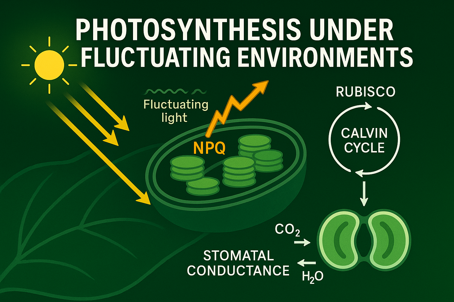
Photosynthesis is one of the earliest physiological processes affected by stress. Whether the stress is drought, heat, salinity, nutrient deficiency, or pathogen attack, the photosynthetic apparatus particularly Photosystem II (PSII) responds almost immediately. Chlorophyll fluorescence arises from excited chlorophyll molecules that cannot use absorbed light energy for photochemistry. As stress disrupts electron transport, energy dissipation pathways change, altering fluorescence signals.
Unlike gas exchange or biochemical assays, chlorophyll fluorescence is non-destructive, rapid, and highly sensitive. Parameters such as maximum quantum efficiency (Fv/Fm), operating efficiency (ΦPSII), non-photochemical quenching (NPQ), and electron transport rates respond dynamically to environmental stress. Importantly, these changes often occur before stomatal closure, growth reduction, or visual damage.
Fluorescence imaging adds a spatial dimension to this information. Instead of providing a single averaged value from a leaf, imaging reveals heterogeneous stress patterns, highlighting specific regions where photosynthetic efficiency begins to decline. This spatial resolution is crucial because stress rarely affects tissues uniformly. Some cells experience photoinhibition earlier, others maintain function longer, and these differences carry valuable physiological information.
The Limitation of Traditional Fluorescence Analysis
Despite its power, conventional chlorophyll fluorescence analysis has limitations. Interpreting fluorescence parameters requires expert knowledge, and subtle stress signatures can be difficult to recognize manually. Fluorescence datasets are also large and multidimensional, containing spatial, temporal, and spectral complexity that exceeds human analytical capacity. Traditional analysis often relies on threshold values or simplified indices, which may overlook early stress signals. For example, a small but consistent spatial decline in ΦPSII across leaf margins may indicate early drought stress, yet remain unnoticed if average values appear normal. Similarly, delayed relaxation of NPQ after light transitions can signal metabolic stress but is difficult to quantify visually. As fluorescence imaging systems became more advanced, generating massive datasets, it became clear that new analytical approaches were needed. This is where artificial intelligence entered plant physiology.
How AI Transforms Chlorophyll Fluorescence Imaging
Artificial intelligence, particularly machine learning and deep learning, excels at identifying patterns in complex datasets. When applied to chlorophyll fluorescence imaging, AI systems learn to recognize stress-specific fluorescence signatures by analyzing thousands of images across different conditions. Deep learning models, such as convolutional neural networks, process fluorescence images pixel by pixel, extracting spatial features, intensity distributions, and temporal dynamics. These models do not rely on predefined thresholds. Instead, they learn directly from data, identifying combinations of fluorescence parameters that consistently correspond to early stress states.
One of the most powerful aspects of AI-driven fluorescence analysis is its ability to detect non-obvious patterns. AI can identify subtle spatial textures, asymmetries, or temporal delays that human observers overlook. For example, slight changes in NPQ distribution across mesophyll regions or delayed recovery of PSII efficiency after light stress can serve as early indicators of heat or drought stress. AI models can also integrate multiple fluorescence parameters simultaneously, creating a holistic stress fingerprint rather than relying on single metrics. This multidimensional approach greatly improves sensitivity and specificity, reducing false positives and improving early detection accuracy.
Early Stress Detection
AI-driven chlorophyll fluorescence imaging has proven effective across a wide range of stress conditions. Under drought stress, AI models detect early reductions in ΦPSII and altered NPQ dynamics long before visible wilting occurs. These changes reflect limitations in electron transport caused by declining CO₂ availability and metabolic constraints. During heat stress, AI identifies spatial photoinhibition patterns associated with thermal damage to PSII and instability of Rubisco activase. These signatures often appear within minutes of heat exposure, allowing rapid stress assessment. In salinity stress, fluorescence imaging combined with AI reveals heterogeneous stress zones linked to ion toxicity and osmotic imbalance. Nutrient deficiencies also produce characteristic fluorescence patterns. Nitrogen limitation alters chlorophyll content and PSII efficiency, while phosphorus deficiency affects energy metabolism and photochemical quenching. AI systems can distinguish between these stresses based on fluorescence signatures alone, even when visual symptoms are identical. In pathogen interactions, AI-driven fluorescence imaging detects localized photosynthetic suppression near infection sites before lesions form. This early warning is particularly valuable for disease management and resistance screening.
From Laboratory to Field
One of the most exciting developments is the scaling of AI-driven chlorophyll fluorescence from controlled environments to real-world agriculture. High-throughput phenotyping platforms now capture fluorescence images of hundreds or thousands of plants simultaneously. AI systems analyze these images in real time, enabling rapid screening of genotypes for stress tolerance. Field-deployable fluorescence systems, combined with drones or robotic platforms, are beginning to emerge. Although field fluorescence imaging is technically challenging due to variable light conditions, AI helps correct for noise, background variation, and environmental fluctuations. This makes large-scale stress monitoring increasingly feasible. In breeding programs, AI-fluorescence systems accelerate selection by identifying stress-resilient genotypes based on early physiological performance rather than final yield alone. This reduces breeding cycles and improves selection accuracy under climate variability.
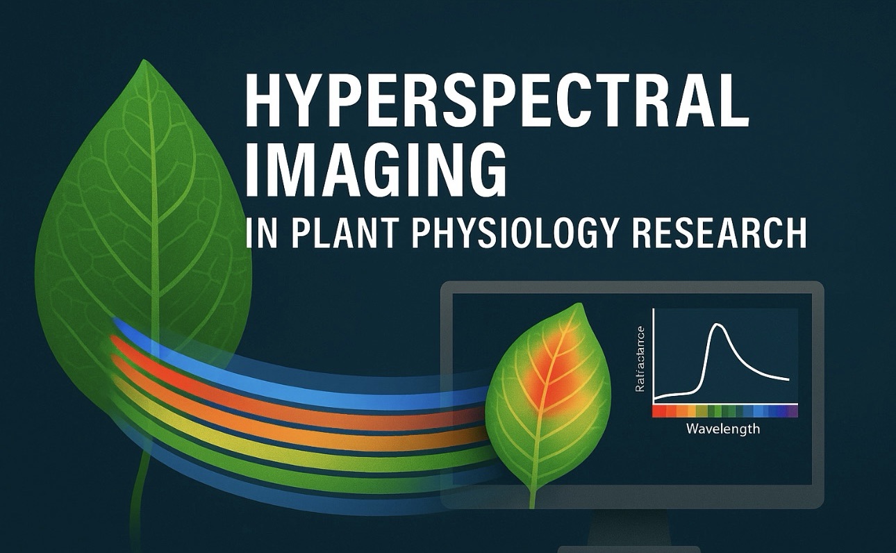
AI-driven chlorophyll fluorescence imaging does not operate in isolation. It integrates seamlessly with other modern plant phenotyping tools such as hyperspectral imaging, thermal imaging, gas exchange, and environmental sensors. When combined, these datasets provide a multi-layered view of plant stress responses. Future systems are likely to use hybrid AI models that combine fluorescence signals with spectral and thermal data, enabling even earlier and more precise stress prediction. Digital plant twins may incorporate fluorescence-derived photosynthetic models to simulate future stress outcomes under changing climate scenarios. As computational power increases and imaging systems become more accessible, AI-driven fluorescence imaging will likely move beyond research settings into routine agricultural practice. Greenhouses, vertical farms, and precision agriculture systems may soon rely on real-time fluorescence monitoring to guide irrigation, nutrient management, and stress mitigation.
Conclusion
AI-driven chlorophyll fluorescence imaging represents a fundamental shift in plant stress detection. Instead of reacting to visible damage, researchers and farmers can now anticipate stress, intervene earlier, and minimize yield losses. By combining the physiological sensitivity of chlorophyll fluorescence with the analytical power of artificial intelligence, plant science has gained a tool that reveals the invisible beginnings of stress. In a world facing increasing climate uncertainty, technologies that allow plants to be monitored at their most vulnerable early stages are not just valuable but they are essential. AI-driven fluorescence imaging brings us closer to truly intelligent plant monitoring systems, where physiology, imaging, and algorithms work together to protect plant productivity and food security.